Peptide Solubility Troubleshooting: Charge, Co-Solvents and Aggregation
Why peptides fail to dissolve: charge and pI-based solvent choice, acids, bases, DMSO and other co-solvents, sonication, aggregation and decision tables.
By the APL Research Team · Updated · First published · 6 min read
Most research peptides dissolve in water within minutes. The ones that do not usually reveal a problem of charge or hydrophobicity, and the fix follows from the sequence rather than from more vigorous mixing. This guide covers how to predict solubility from a sequence, the order in which to escalate solvents, the chemical costs of each option, and how to read a cloudy or gelled solution. Each additive becomes part of the experiment, so the aim is the mildest solvent that works.
Read the sequence first
Three counts predict most outcomes:
- Net charge at the working pH. At pH 7, count +1 for each Lys, Arg and free N-terminus and −1 for each Asp, Glu and free C-terminus. His contributes little at pH 7 but becomes positive below about pH 6. An N-terminal acetyl group removes a positive charge and a C-terminal amide removes a negative one.
- Hydrophobic content. Runs of Leu, Ile, Val, Phe, Trp, Met and Tyr drive self-association, especially when net charge is low.
- Reactive residues. Cys, Met and Trp limit the choice of co-solvent because they oxidise.
The isoelectric point is the pH at which net charge is zero, and solubility usually falls as the pH approaches it. Treat that as a heuristic: in salting-out experiments with several proteins, neither aggregate nor crystal solubility showed a minimum at the pI [1]. Sequence-based predictors can refine the estimate; CamSol now models pH-dependent solubility [2], and CamSol-PTM handles peptides with modified or non-natural residues [3]. How to read peptide sequences covers the notation.
Worked classifications
| Peptide | Sequence | Net charge at pH 7 | Class | First solvent |
|---|---|---|---|---|
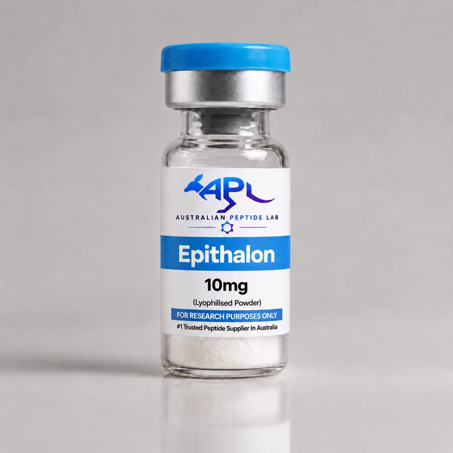
| Epithalon | Ala-Glu-Asp-Gly | −2 | Acidic | Water; dilute ammonium bicarbonate if needed |
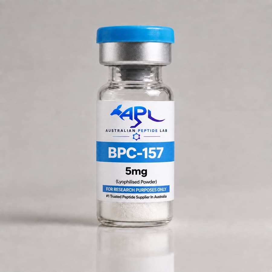
| BPC-157 | GEPPPGKPADDAGLV | −2 | Acidic, proline-rich | Water or neutral buffer |
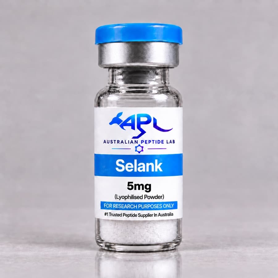
| Selank | Thr-Lys-Pro-Arg-Pro-Gly-Pro | +2 | Basic | Water; dilute acetic acid if needed |
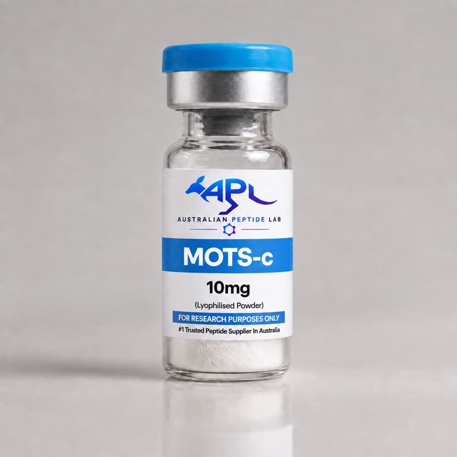
| MOTS-c | MRWQEMGYIFYPRKLR | +3 | Basic, with a hydrophobic aromatic core; two Met, one Trp | Water, then dilute acetic acid; avoid DMSO |
MOTS-c shows why the counts are read together. Its net charge favours aqueous solution, but the central Tyr-Ile-Phe-Tyr stretch adds hydrophobic drive, and the two methionines and one tryptophan make an oxidising co-solvent a poor fallback.
The escalation ladder
Escalate one step at a time, using the smallest volume that could work, because additions accumulate and cannot be taken back. Where the vial holds more peptide than one experiment needs, dissolve it in the minimum volume of the chosen solvent and dilute afterwards.
| Step | Action | Suits | Cost |
|---|---|---|---|
| 1 | Water, gentle swirling, 10–15 minutes at room temperature | Most peptides | None |
| 2 | Move the pH away from the pI: dilute acetic acid for basic peptides, dilute ammonium bicarbonate or ammonia for acidic ones | Charged peptides near their pI | pH-driven chemistry; both additives are volatile and can be removed by lyophilisation |
| 3 | Dissolve in a minimal volume of DMSO, then add the aqueous diluent dropwise while swirling | Hydrophobic peptides with little net charge | Oxidant; cell toxicity; needs a vehicle control |
| 4 | Short bursts of bath sonication with cooling | Persistent particles | Mechanical stress can promote aggregation |
| 5 | Chaotropes such as 6 M guanidine hydrochloride or 8 M urea | Analytical work only | Incompatible with cells and most activity assays |
| 6 | Monomerising solvents (HFIP, or high pH) followed by removal or rapid neutralisation | Amyloidogenic sequences | Specialised methods; validate the product |
The general method for adding diluent to a vial is in the peptide reconstitution guide.
The chemical cost of each option
Alkaline conditions
High pH dissolves acidic peptides but accelerates deamidation and isomerisation. In glucagon held at alkaline pH, deamidation and isomerisation were more common at pH 10 than pH 9; deamidation of Gln3, isomerisation of Asp9 and changes at Asn28 caused marked potency loss, and fibrillation was much greater at pH 9 than pH 10 [4]. Keep exposure to high pH short, dilute or neutralise promptly, and prefer ammonium bicarbonate, which buffers only mildly alkaline, to stronger bases. Deamidation explains the reaction.
Acidic conditions
Dilute acetic acid is the standard aid for basic peptides and has a second advantage: with the decapeptide cetrorelix, acidic media reduced adsorption to vial surfaces by giving the molecules a repulsive charge [5]. Stronger or prolonged acid exposure risks backbone cleavage at Asp-Pro bonds, so acid is a solvent aid rather than a storage medium.
DMSO
DMSO dissolves almost anything, which hides its drawbacks. It is an oxidant used deliberately in peptide synthesis to form disulfide bonds [6], so it is a poor choice for peptides with free cysteine and a questionable one for methionine-rich sequences. In cells, DMSO above 1% (v/v) induced death in a retinal neuronal line, and the authors recommended computing absolute final DMSO concentrations and running untreated as well as vehicle controls [7]. Concentrations as low as 0.0008–0.004% changed signalling proteins across eight lung cancer cell lines, heterogeneously from line to line [8].
Sonication
Sonication breaks up particles and is part of a validated method for preparing seedless amyloid-β monomer, combined with dissolution above pH 12 and rapid freezing [9]. The same energy can drive aggregation: under ultrasonication, transthyretin formed mature amyloid fibrils at neutral pH that it did not form when left quiescent [10]. Use short bursts in a cooled bath and inspect the result between them.
Aggregation-prone peptides
Some peptides dissolve and then aggregate. For amyloid-β, the formation of oligomers, fibrils and larger aggregates was governed by time, concentration, temperature, pH, ionic strength and peptide species [11], so the same powder can give different solutions depending on handling. Laboratories that need defined starting material first treat the dry peptide with a strong solvent to erase any "structural history" that could seed aggregation [12], and a recent comparison found that mild solvent treatments often failed to give consistently monomeric preparations [9]. For these sequences, dissolve at stock concentration in the favourable solvent, dilute into buffer at the last step, and use the solution promptly. Peptide aggregation summarises the mechanisms.
Reading a cloudy or gelled solution
| Observation | Likely cause | Quick test | Action |
|---|---|---|---|
| Haze that clears when pH is shifted | pH near the pI | Add a small volume of dilute acid or base to a test portion | Adjust pH of the stock |
| Gel or viscous solution | Self-assembly | Dilute a portion twofold | Lower the concentration; consider a monomerising solvent |
| Particles that settle | Undissolved peptide or aggregates | Spin down and measure the supernatant | Escalate one step |
| Precipitate when stock meets buffer or medium | Solvent shock or salting-out | Add stock slowly to the full volume with mixing | Lower stock concentration or add the buffer salts last |
| Clear, but measured concentration low | Adsorption or an undissolved film | Rinse the vial and measure | Low-binding tubes; acidify basic peptides |
| Cloudiness appearing days later | Aggregation or microbial growth | Inspect under a microscope | Discard; review sterile technique |
Filtering haze away also removes peptide, so a filtrate's concentration is no longer the one calculated.
Co-solvents compared
| Co-solvent | Typical use | Removable by lyophilisation | Cell assays | Cautions |
|---|---|---|---|---|
| Dilute acetic acid | Basic peptides | Yes | After dilution | Low pH during dissolution |
| Dilute ammonium bicarbonate | Acidic peptides | Yes | After dilution | Mildly alkaline; deamidation |
| DMSO | Hydrophobic, low-charge peptides | No | Low final percentage, with vehicle control | Oxidant; signalling effects |
| Acetonitrile | HPLC and MS samples | Yes | No | Analytical use only |
| HFIP | Monomerising amyloidogenic peptides | Evaporated before use | Only after complete removal | Corrosive, volatile |
| Guanidine hydrochloride or urea | Denaturing analytical work | No | No | Urea solutions can carbamylate amines |
For the bench
- Record the final solvent composition of every stock; it defines the vehicle control. Bacteriostatic water vs sterile water shows how to keep the vehicle constant across a concentration series.
- Recalculate concentration after any additions; the molarity calculator and dilution calculator handle the volumes, and peptide molarity calculations covers net peptide content.
- Note a peptide that dissolves less readily than earlier vials of the same lot; it may signal aggregation during storage, covered in the peptide storage guide.
Frequently asked questions
Why does a peptide fail to dissolve in water?
Usually because the solution pH sits close to its isoelectric point, so the molecules carry little net charge to keep them apart, or because a run of hydrophobic residues drives self-association. Less often the peptide has already aggregated in the vial. Counting charged residues predicts most cases, and sequence-based predictors such as CamSol estimate how solubility changes with pH [2].
Is DMSO acceptable for peptide stocks used in cell assays?
It can be, with limits. DMSO above 1% (v/v) killed a retinal neuronal cell line [7], and even 0.0008–0.004% altered the expression or activation of signalling proteins in lung cancer cell lines [8]. Use the least DMSO that dissolves the peptide, report the final percentage, and include a vehicle control at the same concentration. Avoid it for peptides with free cysteine, because DMSO is an oxidant [6].
Should a stubborn peptide solution be sonicated?
Briefly, as a later step, and with the result checked. Sonication breaks up particles, and a high-pH dissolution method for amyloid-β includes it [9]. Mechanical stress can also push susceptible molecules towards aggregation: ultrasonication produced mature transthyretin amyloid fibrils at neutral pH where quiescent incubation did not [10]. Use short bursts in a cooled bath and inspect between them.
Is a peptide least soluble at its isoelectric point?
As a rule of thumb, yes: at the pI the net charge is zero and electrostatic repulsion is weakest. It is not a law. In salting-out experiments with several proteins, neither aggregate nor crystal solubility had a minimum at the isoelectric point [1]. Treat the pI as a guide to which direction to move the pH, then test.
Can solubility be predicted before opening the vial?
Roughly. Net charge at the working pH and the proportion of hydrophobic residues give a first estimate, and how to read peptide sequences shows how to count them. Computational tools go further: CamSol predicts pH-dependent solubility from sequence [2], and CamSol-PTM extends predictions to peptides containing modified or non-natural amino acids [3].
References
- 1.Dumetz AC, Chockla AM, Kaler EW, et al. Effects of pH on protein-protein interactions and implications for protein phase behavior. Biochim Biophys Acta. 2008. PubMed 18258214
- 2.Oeller M, Kang R, Bell R, et al. Sequence-based prediction of pH-dependent protein solubility using CamSol. Brief Bioinform. 2023. PubMed 36719110
- 3.Oeller M, Kang RJD, Bolt HL, et al. Sequence-based prediction of the intrinsic solubility of peptides containing non-natural amino acids. Nat Commun. 2023. PubMed 37978172
- 4.Caputo N, Castle JR, Bergstrom CP, et al. Mechanisms of glucagon degradation at alkaline pH. Peptides. 2013. PubMed 23651991
- 5.Grohganz H, Rischer M, Brandl M. Adsorption of the decapeptide Cetrorelix depends both on the composition of dissolution medium and the type of solid surface. Eur J Pharm Sci. 2004. PubMed 14757490
- 6.Tamamura H, Otaka A, Nakamura J, et al. Disulfide bond-forming reaction using a dimethyl sulfoxide/aqueous HCl system and its application to regioselective two disulfide bond formation. Int J Pept Protein Res. 1995. PubMed 7601603
- 7.Galvao J, Davis B, Tilley M, et al. Unexpected low-dose toxicity of the universal solvent DMSO. FASEB J. 2014. PubMed 24327606
- 8.Baldelli E, Subramanian M, Alsubaie AM, et al. Heterogeneous Off-Target Effects of Ultra-Low Dose Dimethyl Sulfoxide (DMSO) on Targetable Signaling Events in Lung Cancer In Vitro Models. Int J Mol Sci. 2021. PubMed 33802212
- 9.Taylor AIP, Davis PJ, Aubrey LD, et al. Simple, Reliable Protocol for High-Yield Solubilization of Seedless Amyloid-β Monomer. ACS Chem Neurosci. 2023. PubMed 36512740
- 10.Yuzu K, Yamamoto N, Matsumura M, et al. Ultrasonication-induced in vitro formation of transthyretin mature amyloid fibrils at neutral pH. Protein Sci. 2026. PubMed 41793160
- 11.Stine WB Jr, Dahlgren KN, Krafft GA, et al. In vitro characterization of conditions for amyloid-beta peptide oligomerization and fibrillogenesis. J Biol Chem. 2003. PubMed 12499373
- 12.Stine WB, Jungbauer L, Yu C, et al. Preparing synthetic Aβ in different aggregation states. Methods Mol Biol. 2011. PubMed 20967580
This article summarises published research for educational purposes. It is not medical advice. Compounds sold by Australian Peptide Lab are research reagents for in-vitro laboratory use only, not for human or veterinary use.