Sterile Technique for Peptide Solutions: Aseptic Bench Practice, Filtration and Records
Aseptic technique for peptide solutions: clean benches vs biosafety cabinets, routine, sterile filtration losses, endotoxin, equipment and records.
By the APL Research Team · Updated · First published · 6 min read
Aseptic technique keeps a sterile solution sterile from the moment it is prepared until it reaches the assay; it cannot make a contaminated solution clean again. For peptide work the stakes are specific: microbial growth consumes and degrades peptide, endotoxin activates immune cells at trace levels, and mycoplasma alters the cells the peptide is tested on. This guide covers the workspace, the routine, sterile filtration and its losses, endotoxin, contamination monitoring, the equipment a small peptide bench needs, and the records that make results traceable.
Where contamination comes from
Aseptic technique is broader than sterile consumables: it spans environmental control, personal hygiene, sterilisation of equipment and media, and the quality-control checks that confirm the work was clean [1]. Contamination enters cell-culture work from the operator and the laboratory environment, from other cells handled in the same space, and from reagents [2].
| Source | Typical route | Control |
|---|---|---|
| Operator | Skin, hair, breath, talking over open vessels | Gloves, cuffed lab coat, slow movements inside a cabinet |
| Environment | Air currents, dust, traffic past the bench | Cabinet work, closed doors, low-traffic location |
| Reagents | Diluents, buffers, serum, peptide stocks | Sterile or filtered stocks, single-use aliquots, recorded lot numbers |
| Other cultures | Shared reagents, simultaneous handling | One cell line at a time, separate reagent sets |
| Equipment | Pipettes, water baths, incubators | Filtered tips, regular cleaning and maintenance |
Cell lines are also frequently misidentified or contaminated by other cells, bacteria, fungi, yeast, viruses or chemicals, which is why biosafety cabinets and protective equipment are standard [3].
Choosing the workspace
Most cell-culture work uses either a horizontal laminar-flow clean bench or a vertical-flow biosafety cabinet [1]. They are not interchangeable:
| Workspace | Protects the solution | Protects the operator | Suits |
|---|---|---|---|
| Open bench | Only if containers stay closed | No | Septum entry into sealed vials; non-sterile preparation |
| Horizontal laminar-flow clean bench | Yes | No; air flows towards the operator | Sterile preparation of non-hazardous solutions, such as diluting peptide stocks for biochemical assays |
| Class II biosafety cabinet | Yes | Yes | Cell culture and any work with biological material |
A cabinet protects only when used well. In aerosol challenge tests on a Class II cabinet, inflow velocity, the size of the access opening, the type and pace of the operator's movements and the position of the hands all influenced containment, and measuring airflow alone did not guarantee protection [4]. In a later comparison of operator protection, one Class II model passed every test, including those with fans, opening doors and people walking past, while a second gave poor protection whenever those interfering factors were present and failed the standard test at the lowest airflow [5]. Cabinets also drift out of specification: of 360 Class II cabinets tested in Beijing hospitals between 2018 and 2023, 70.4% met all performance criteria, with inflow and downflow velocity among the indicators most often failing [6]. Certification is worth checking before relying on one.
The routine
- Switch the cabinet on for the purge time its manufacturer specifies, then wipe the work surface with 70% ethanol.
- Load only what the task needs, keep the front and rear grilles clear, and arrange items from clean to used.
- Wear nitrile gloves and a cuffed lab coat; spray gloves with 70% ethanol after touching anything outside the cabinet.
- Work well inside the cabinet with slow, deliberate movements, and never pass a hand or sleeve over an open vessel.
- Wipe every vial septum and bottle neck with 70% alcohol and let it dry completely before entry; wet alcohol carried in on a needle or tip defeats the purpose.
- Use each sterile tip, pipette or needle once, and never return unused liquid to a stock container.
- Close everything, wipe down, and dispose of sharps and biological waste through the correct streams.
For sealed septum vials, enter through the centre of the stopper with a new sterile needle each time, and vary the entry point slightly so the septum is not cored. The peptide reconstitution guide covers how diluent goes in; the same routine applies to every later withdrawal.
Sterile filtration of peptide solutions
Filtration is needed when a peptide stock destined for cell culture was made from components that were not sterile, such as a co-solvent, a buffer prepared on the open bench, or a vial opened outside a cabinet. A 0.22 µm or 0.2 µm sterilising-grade membrane is standard, with three caveats.
Retention is probabilistic. In a challenge study, Ralstonia pickettii cells that shrank during exposure to a drug solution penetrated 0.2 µm nylon and 0.22 µm PVDF sterilising-grade filters in some tests, while 0.1 µm filters qualified with the mycoplasma Acholeplasma laidlawii consistently gave sterile filtrate [7]. Where mycoplasma is a concern, as in preparing media components, 0.1 µm filters give an extra margin.
Filters remove peptide as well as organisms. Syringe filter units with glass-microfibre prefilters lost 20–80% of a protein by adsorption, whereas membrane-only units lost 0–20%; filters also shed particles, from essentially none to more than 100,000 per mL, that accelerated aggregation when the filtrate was agitated [8]. With infusion filters, adsorption depended mainly on the filter material and the diluent, with strongly negatively charged membranes adsorbing most in glucose solution [9].
Filtration does not remove endotoxin or dissolved contaminants. It removes organisms, not what they leave behind.
In practice: choose a membrane-only, low-protein-binding filter sized to the volume, filter while the stock is concentrated so adsorption takes a smaller fraction, check the membrane's compatibility with any organic co-solvent, avoid shaking the filtrate, and measure the concentration after filtration. The sterile filtration entry summarises the terms.
Endotoxin
Sterility and endotoxin are separate specifications. When reagents used in microglia research were screened with the Limulus amoebocyte lysate (LAL) assay, significant lipopolysaccharide contamination was found in a number of commercial-grade proteins, phospholipids and synthetic peptide preparations, but not in pharmaceutical-grade recombinant proteins; polymyxin B, which neutralises lipopolysaccharide, suppressed the microglia-activating effect of several commercial preparations [10]. For macrophage, microglia, dendritic-cell or other inflammation assays, test peptide stocks and diluents by LAL where the result matters, use endotoxin-specified water and plasticware, and include a polymyxin B control arm. Endotoxin explains the chemistry, and bacteriostatic water vs sterile water covers the choice of diluent.
Watching for contamination
Bacterial and fungal contamination usually announces itself: turbidity, a rapid colour shift in the medium's pH indicator, films or floating colonies. Mycoplasma does not. A survey of 9,395 rodent and primate samples in NCBI's RNA-seq archive found 11% of 884 study series contaminated, with mycoplasma read counts associated with changes in host gene expression [11]. When the US National Center for Advancing Translational Sciences introduced routine testing, its initial mycoplasma contamination rate was above 10%, and short tandem repeat profiling of 186 cell lines identified five misidentified lines, all received from external laboratories [12].
When contamination is found, discarding the affected culture may be enough; if the problem is wider, discard all contaminated cultures and any media opened during the period, inspect and clean equipment, and review procedures before resuming [2]. Quarantine and test new cell lines before they enter general use.
Equipment for a small peptide bench
| Function | Item | Notes |
|---|---|---|
| Storage | −20 °C manual-defrost freezer; 2–8 °C refrigerator | Min–max thermometer or data logger in each |
| Storage | Desiccator or sealed box with desiccant | For lyophilised vials in use |
| Aseptic work | Certified Class II cabinet or laminar-flow clean bench | Match the workspace to the task |
| Aseptic work | 70% ethanol, lint-free wipes, alcohol swabs, nitrile gloves, cuffed lab coat | Consumed daily |
| Liquid handling | Calibrated pipettes with filtered tips; sterile serological pipettes | Calibration dates recorded |
| Liquid handling | Sterile single-use syringes and needles; sharps container | For septum vials |
| Containers | Low-binding polypropylene tubes; screw-cap cryotubes; vial racks | Low-binding matters for dilute peptide solutions |
| Filtration | 0.22 µm membrane-only, low-binding syringe filters | 0.1 µm where mycoplasma is a concern |
| Measurement | Micro-volume UV spectrophotometer; pH meter | Concentration and pH checks |
| Records | Bound notebook or electronic system; cryo-rated labels | See below |
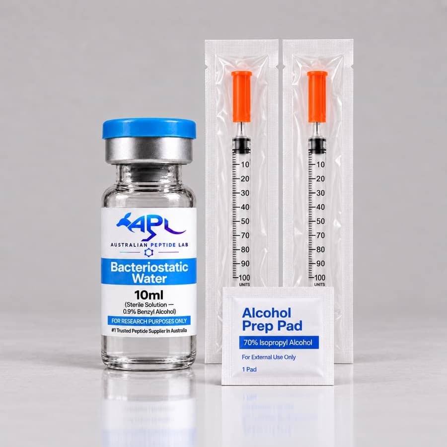
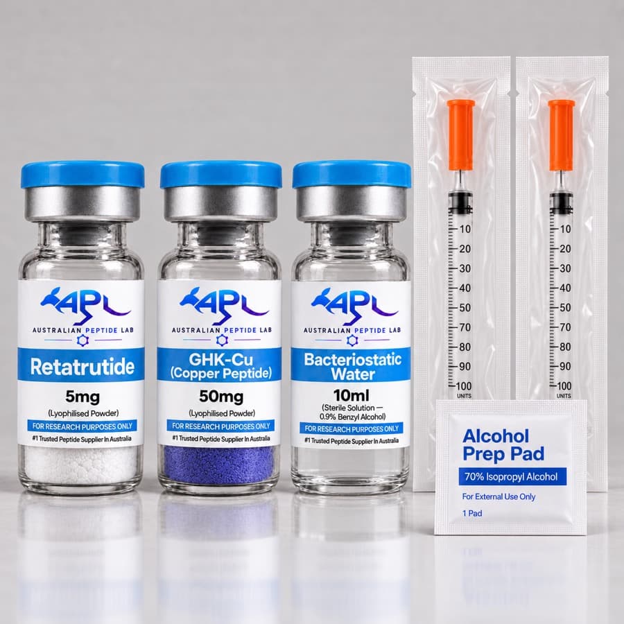
Our Research Starter Kit covers the diluent, syringes and swabs, bacteriostatic water is available separately, and other consumables are in lab supplies.
Record keeping
A result that cannot be traced to a lot, a preparation date and a storage history cannot be properly interpreted later. Keep five linked records:
| Record | Contents |
|---|---|
| Receipt log | Lot number, date received, packaging condition, cake appearance, storage location |
| Preparation record | Date, operator, diluent and its lot, volume, concentration and whether net peptide content was applied, final solvent composition, filtration step |
| Aliquot register | Aliquot IDs, volumes, freezer positions, thaw dates, freeze–thaw count |
| Equipment log | Freezer and refrigerator temperatures, cabinet certification dates, pipette calibration |
| Deviation log | Temperature excursions, contamination events, actions taken |
The lot number is the thread that joins them to the batch documentation; understanding certificates of analysis explains what that documentation contains, and the peptide storage guide covers the storage side of the log.
Frequently asked questions
Is a laminar-flow clean bench the same as a biosafety cabinet?
No. A horizontal laminar-flow clean bench blows filtered air across the work towards the operator, protecting the product but not the person. A Class II biosafety cabinet draws air in at the front and sends filtered air down over the work, protecting both. Both are used for cell-culture work [1]; anything involving cells or other biological material belongs in the cabinet.
Does 0.22 µm filtration guarantee a sterile solution?
It is the standard sterilising step, but retention is probabilistic. In one challenge study, Ralstonia pickettii that had shrunk in a drug solution passed some 0.2 and 0.22 µm sterilising-grade filters, while 0.1 µm filters qualified with a mycoplasma gave sterile filtrate [7]. Filtration also leaves endotoxin and dissolved contaminants behind in the solution, so it complements aseptic technique rather than replacing it.
How much peptide is lost in a syringe filter?
It varies with the filter and the solution. For a protein filtered through syringe units, those with glass-microfibre prefilters lost 20–80% by adsorption and membrane-only units 0–20% [8]. Adsorption to infusion filters depended mainly on the membrane material and the diluent [9]. Filter while the solution is concentrated, choose a membrane-only low-binding unit, and measure the concentration afterwards.
Does bacteriostatic water make aseptic technique unnecessary?
No. Its 0.9% benzyl alcohol limits growth of small numbers of organisms introduced on entry, but it does not sterilise a heavily contaminated vial or remove endotoxin. Swabbing, sterile consumables and single-use aliquots still do most of the work. See what is bacteriostatic water for what the preservative can and cannot do.
Why test cell cultures for mycoplasma if they look healthy?
Because mycoplasma does not cloud the medium. A survey of public RNA-seq data found contamination in 11% of 884 study series [11], and a translational research centre found an initial contamination rate above 10% when it introduced routine testing [12]. Peptide effects measured in infected cultures may reflect the infection rather than the peptide.
References
- 1.Coté RJ. Aseptic technique for cell culture. Curr Protoc Cell Biol. 2001. PubMed 18228291
- 2.Stacey GN. Cell culture contamination. Methods Mol Biol. 2011. PubMed 21516399
- 3.Weiskirchen S, Schröder SK, Buhl EM, et al. A Beginner's Guide to Cell Culture: Practical Advice for Preventing Needless Problems. Cells. 2023. PubMed 36899818
- 4.Macher JM, First MW. Effects of airflow rates and operator activity on containment of bacterial aerosols in a class II safety cabinet. Appl Environ Microbiol. 1984. PubMed 6437327
- 5.Parks S, Hookway H, Kojima K, et al. The Impact of Air Inflow and Interfering Factors on the Performance of Microbiological Safety Cabinets. Appl Biosaf. 2022. PubMed 36032323
- 6.Song T, Yin S, Liu Z, et al. Performance evaluation of biological safety cabinets: a real-world analysis. Front Bioeng Biotechnol. 2026. PubMed 42212170
- 7.Sundaram S, Auriemma M, Howard G Jr, et al. Application of membrane filtration for removal of diminutive bioburden organisms in pharmaceutical products and processes. PDA J Pharm Sci Technol. 1999. PubMed 10754712
- 8.Liu L, Randolph TW, Carpenter JF. Particles shed from syringe filters and their effects on agitation-induced protein aggregation. J Pharm Sci. 2012. PubMed 22674153
- 9.Besheer A. Protein Adsorption to In-Line Filters of Intravenous Administration Sets. J Pharm Sci. 2017. PubMed 28559043
- 10.Weinstein JR, Swarts S, Bishop C, et al. Lipopolysaccharide is a frequent and significant contaminant in microglia-activating factors. Glia. 2008. PubMed 17910052
- 11.Olarerin-George AO, Hogenesch JB. Assessing the prevalence of mycoplasma contamination in cell culture via a survey of NCBI's RNA-seq archive. Nucleic Acids Res. 2015. PubMed 25712092
- 12.Roth JS, Lee TD, Cheff DM, et al. Keeping It Clean: The Cell Culture Quality Control Experience at the National Center for Advancing Translational Sciences. SLAS Discov. 2020. PubMed 32233736
This article summarises published research for educational purposes. It is not medical advice. Compounds sold by Australian Peptide Lab are research reagents for in-vitro laboratory use only, not for human or veterinary use.